Flow Cytometry
Georgia Cancer Center Flow Cytometry Shared Resource was established in 2006 and provides access to contemporary flow cytometers, support equipment, and associated software and services. The core offers the ability to perform almost every published flow cytometry protocol.
Flow Cytometry Resource is equipped with seven flow cytometers categorized into two types. The first type are analyzer flow cytometers that are typically operated by investigators themselves. The second type are cell sorter flow cytometers that are provided as a service to investigators.
Request training or sorting appointments.
There is a minimum one hour charge for all assisted use which includes analyzers and sorters.
Jump to: Links Bulletin Board Publication Requirements Analyzers Sorters Support Equipment Our Team
Links

Bulletin Board
Recent Events
Summer Research Experience
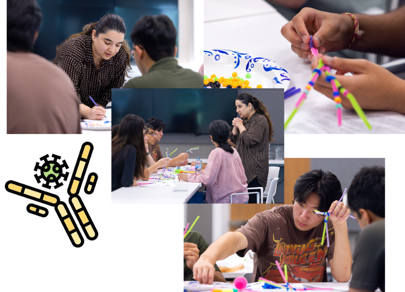
Antibody construction- Dr Ishita Tandon
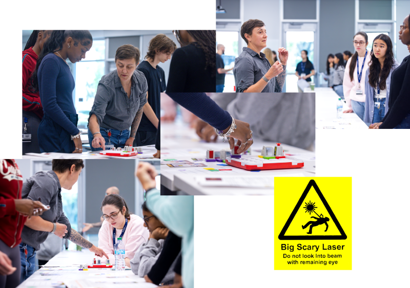
Optics- Dr Rebekah Tritz
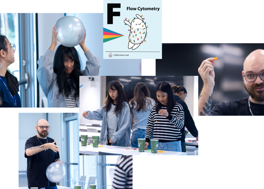
Basics of Sorting- David Hansen
A morning of dry lab demos with the high school students taking part in the GCC Summer Research Experience! Students learned about antibody construction, the optical layout of cytometers and the basics of cell sorting.
Publication Requirements
To cite or acknowledge the use of our core: Georgia Cancer Center Flow and Mass Cytometry Core Facility (RRID: SCR_025747)
All users of the Georgia Cancer Center Flow Cytometry Shared Resource should acknowledge the facility in all publications resulting from work performed in the resource.
For details on acknowledgment requirements and types please go here.
Analyzers
| Location CN 4158C |
|||
| Conventional cytometers
The flow core has four conventional analyzers with 4 - 5 lasers |
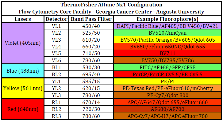
ThermoFisher AttuneNxT
4-lasers, dual focusing capabilities and both tube and plate options. Please click here for machine configuration. SOP found here Assisted/ training cost: $50/hr Unassisted cost: $25/hr |
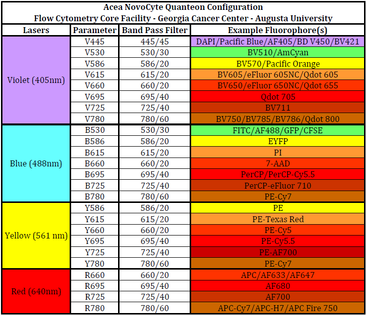
Agilent Quanteon (1 and 2)
4-lasers, high throughput, 25 detectors and both tube and plate options. Please click here for machine configuration. SOP found here Troubleshooting guide found here Assisted/ training cost: $75/hr Unassisted cost: $45/hr |
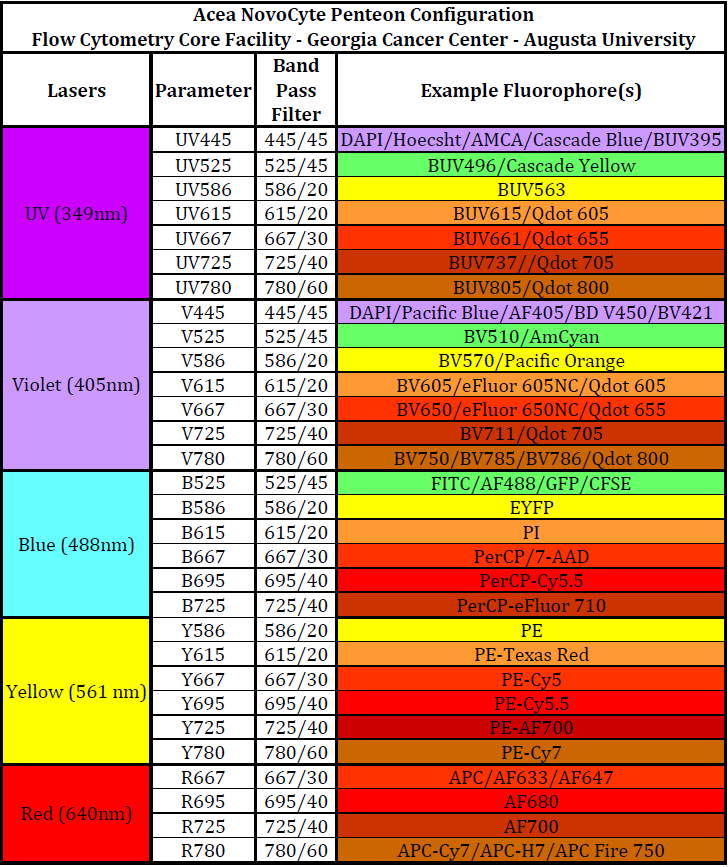
Agilent Penteon
5-lasers, high throughput, 30 detectors and both tube and plate options. Please click here for machine configuration. Assisted/ training cost: $75/hr Unassisted cost: $45/hr |
|
Spectral Cytometer |
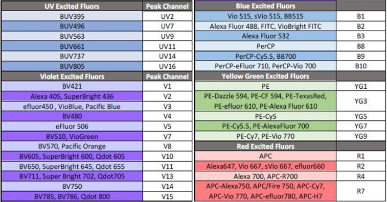
Cytek Aurora
5-lasers, both tube and plate options, 64 detectors, autofluorescence extraction and 40+ color panel capabilities. Please click here for peak channel information. SOP found here Troubleshooting guide found here Assisted/ training cost: $75/hr Unassisted cost: $50/hr |
Cytek EVO SOP found here Troubleshooting guide here Assisted/ training cost: $75/hr Unassisted cost: $50/hr
|
|
Sorters
Miltenyi - MACs Quant Tyto
Location CN 4158C
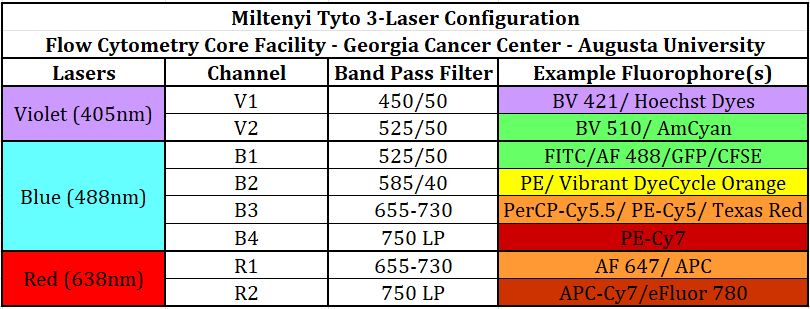
- Microfluidic sorter - perfect for extremely sensitive samples or those requiring sterile sort conditions. This machine has 3 lasers and 8 fluorescent channels. Please click here for machine configuration.
Assisted and unassisted cost:
- regular cartridge $100/ cartridge
- high-speed cartridge $150/ cartridge
ThermoFisher - Bigfoot
Location CN 4146C
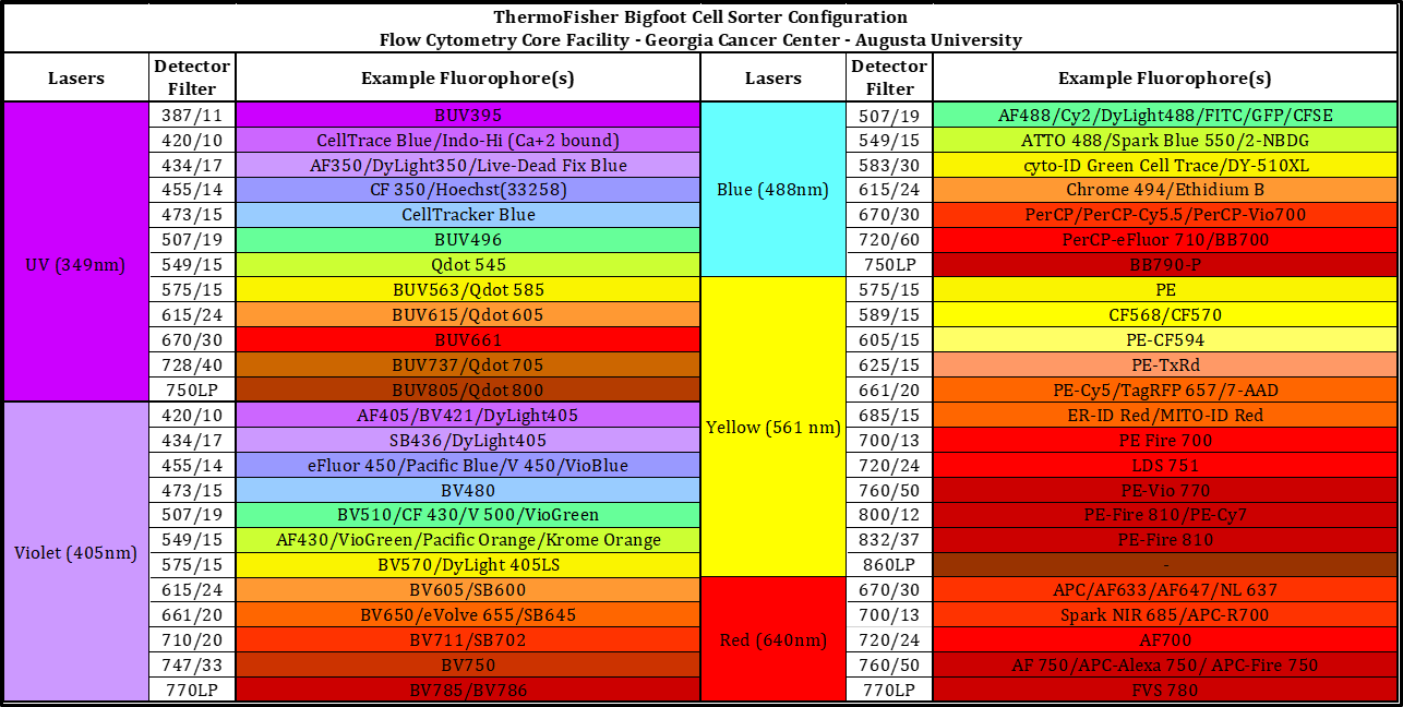
Cytek CS
Location CN 4146C
- Both conventional and spectral sorting capabilities. The CS matches our Aurora and EVO analyzers in configuration and software. This cuvette sorter can sort 6 populations at a time into a variety of collection tubes. Please click here for machine configuration and here for a sort request.
- Assisted ONLY: $90/hr
Support Equipment
Location CN 4158D
Computer Analysis Workstations
Analysis software available: NovoExpress, FlowJo, SpectroFlo, OMIQ, Attune Nxt and CytExpert
Facility users: no charge
Non-facility users: $10/hr
Consultation
no charge
Technical Assistance
$75/hr - users will be notified before initiation of charged time
Our Team