Selner's Rhinolaryngoscopy Online
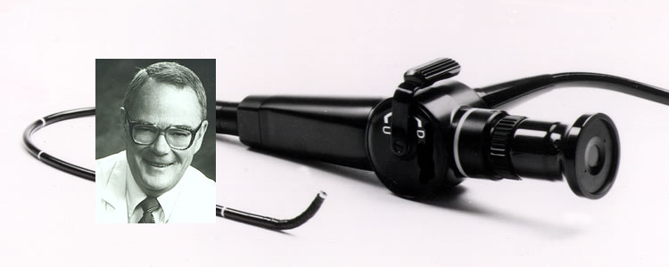
John C. Selner, MD
William K. Dolen, MD
Bryan Spofford, MD
Jerald W. Koepke, MD
Chapter 4. UPPER AIRWAY PATHOLOGY
Too often, a patient complaining of nasal congestion or other upper airway symptoms will be placed on decongestant therapy, antibiotics, or allergen immunotherapy when symptoms are largely due to anatomic obstruction of the nasal passage or other conditions primarily managed by surgery. By routine examination, it is difficult to diagnose posterior deviation of the nasal septum or to detect nasopharyngeal obstruction by a choanal polyp or hypertrophied adenoidal tissue. The fiberoptic rhinolaryngoscope is a convenient tool for direct inspection of nearly all portions the upper airway. This chapter will summarize pathology of the upper airway as it might be encountered in fiberoptic endoscopy; a complete discussion of upper airway pathology may be found in any textbook of otolaryngology.
MUCOSA
Findings and pathology evident on fiberoptic endoscopy of the upper airway
| Region | Findings |
| Mucosa |
|
Nasal Cavity
| Turbinates |
|
| Septum |
|
| Polyps |
|
| Paranasal sinuses |
|
Nasopharynx and Superior Oropharynx
| Choana | Atresia or stenosis (unilateral or bilateral) |
| Torus, eustachian tube orifice |
|
| Adenoids |
|
| Pharyngeal wall |
|
| Malignancies |
|
Oropharynx, Hypopharynx, Larynx
| Posterior tongue |
|
| Lingual tonsils |
|
| Epiglottis |
|
| Glottis |
|
| Arytenoids |
|
| Vocal cords |
|
In health, the upper airway mucosa are moist and reddish-pink in color; the nasal mucosa shrink easily when topical decongestants are applied. Dryness of the mucosa may accompany various systemic diseases (such as "sicca syndrome"), may result from living in a low humidity environment, or may be a feature of atrophic rhinitis or rhinitis sicca. Allergic rhinitis classically produces a bluish discoloration of the nasal mucosa in association with clear rhinorrhea. So-called "cobblestoning" of the mucosa is a sign of chronic inflammation.
When an exudate is present, its consistency and clarity may be evaluated by rhinoscopy. Presence of a purulent exudate draining from a paranasal sinus orifice would virtually be pathognomonic of sinusitis, although this observation does not permit differentiation of bacterial, viral, or inflammatory etiologies.
NASAL CAVITY
Turbinates
Hypertrophy of the nasal turbinates may occur secondary to chronic inflammation of the nasal mucosa, especially that of the paranasal sinuses, or in cases of septal deviation as a compensatory mechanism in the nasal passage opposite to the obstructed side. Turbinate hypertrophy may also occur without obvious underlying cause. Complete or partial turbinectomy may have been performed in an attempt to relieve obstructive symptoms or as an adjunct in more complicated nasal surgery.
Horizontal or sagittal clefting of a turbinate is a normal anatomic variant which may be confused with polypoidal degeneration. It may also be difficult to distinguish polypoidal degeneration of a turbinate from polyps which are emanating from the anterior group of ethmoid air cells and entering the nose via the middle meatus. In either case, manipulation of the suspected polyps under direct visualization may be useful.
When misplaced ethmoid air cells localize in the concha of a middle turbinate, the resulting structure is a concha bullosa, also termed bullous degeneration of the turbinate, or, more simply, bullous turbinate. A concha bullosa may produce significant obstruction of the nasal airway.
Septum
The nasal septum is not normally a perfectly straight midline structure. In normals, one side of the nasal cavity is larger than the other, but differences are not perceived. More prominent septal deviation will result in obstructive symptoms, and complex deviation will produce bilateral obstruction. Occasionally, severe deviation of the septum will not even permit passage of the rhinoscope. Septal deviation may compress the nasal turbinates and cause facial pain.
A variety of pathological conditions can result in perforation of the septum. Infection of a posttraumatic septal hematoma will cause perforation. Septal perforation may also be associated with abuse of cocaine or the topical nasal decongestants. Patients with septal perforation may complain of nasal congestion, intermittent bleeding, and a whistling sound with nasal breathing.
A septal spur is a displacement of the perpendicular plate of the ethmoid bone and the quadrangular septal cartilage into the nasal cavity. If this spur impinges on adjacent mucosal tissues, facial pain will result. Occasionally, the maxilla will form a shelf, or ridge, on which the cartilaginous portion of the septum rests. This maxillary ridge is a common finding, but may be sufficiently large to cause symptomatic obstruction.
Nasal polyps
Polyps can originate from degeneration of the mucosa of any structure in the nasal cavity or may extend from any of the sinuses, but the vast majority have their origin in the ethmoid air cells. Polyps from the anterior and middle ethmoid air cells enter the nasal cavity from the meatus of the middle turbinate. They are usually easy to visualize on routine anterior examination of the nose, may extend anteriorly and can produce total nasal obstruction. It is not possible to detect posterior ethmoid polyps on routine examination unless they have reached huge proportions.
Polyps characteristically slightly yellow or translucent, smooth avascular structures. They can often be moved with a cotton tipped applicator so that their origin may more clearly be defined. Squamous metaplasia results as a consequence of dry air inducing keratinization of the mucosa covering the polyp.
Polyps located in the middle meatus have their origin from anterior ethmoid or maxillary sinuses. Polyps extending from the sphenoethmoidal recess originate from the sphenoid or posterior ethmoid sinuses.
Neoplasms
Malignancy in the nose is an uncommon finding, but complaints as common as pain, burning, or rhinorrhea can be the initial symptoms of malignant degeneration. Description of the various malignancies of the upper airway is beyond the scope of this text. Any lesion suspicious for malignancy should be evaluated by an otolaryngologist.
Nasal surgery
It is often helpful to examine the results of surgical intervention. Commonly encountered procedures include repair of deviation or perforation of the septum, placement of antral windows, turbinectomy, and exteriorization, or marsupialization, of the ethmoid and sphenoid sinuses.
Paranasal sinus surgery
The maxillary sinus is also called the antrum of Highmore. The establishment of drainage by creation of a dependent antral window in the inferior meatus is a common surgical procedure. With the fiberoptic endoscope, one may assess patency of the antral window and inspect the surrounding mucosa for signs of inflammation or polypoidal growth. It may be possible to insert the endoscope into the maxillary sinus to examine the mucosa.
Ethmoidectomy and sphenoidectomy are performed to establish drainage from and provide aeration to these areas in order to treat chronic inflammation and infection At times, extensive surgery is performed.
NASOPHARYNX, SUPERIOR OROPHARYNX
Choana
The choana marks the beginning of the nasopharynx. Although complete bilateral choanal atresia almost always presents at birth, unilateral atresia or choanal stenosis might not be diagnosed until adulthood.
Torus tubarius
The torus is a partial ring of tissue protecting the ostium of the eustachian tube. A variety of conditions involving the torus could produce symptoms and signs of eustachian tube dysfunction. A cyst of the torus blocking the eustachian tube orifice would be evident on fiberoptic endoscopy, as would lymphoid infiltration from chronic inflammation, and edema of the torus. Since adenoidectomy is often done by blind curettage, damage to the eustachian tube and torus with resulting formation of scar tissue and adhesions could lead to eustachian tube dysfunction.
Rosenmueller s fossa
This vertical cleft, a potential space between the posterior lip of the torus tubarius and the adenoidal pad is of considerable importance because many of the insidious malignancies of the pharynx have their origin here. It is impossible to examine this space without direct visualization.
Adenoid
The adenoid is a primary lymph node of first line defense for inflammation involving the upper airway. The torus tubarius and orifice of the tympanopharyngeal duct (eustachian tube) are in close proximity to the adenoidal pad. Function of the eustachian tube may not infrequently be compromised by adenoidal hypertrophy.
Hypertrophy of the adenoids is not a rare cause of nasal obstruction in children; in extreme cases, anterior herniation of adenoidal tissue through the choanae may result in total obstruction. In addition to producing discomfort, obstruction may result in hyponasal speech, other variations in voice quality, orthodontic difficulties due to abnormal development of facial bones (adenoidal facies), and sleep disturbances. A juvenile nasopharyngeal angiofibroma may mimic the obstruction of adenoidal hypertrophy and may be associated with recurrent epistaxis.
Anterior, rhinorrhea may be associated with severe adenoidal hypertrophy or other obstructing lesions of the nasopharynx. Chronic adenoiditis may result in posterior rhinorrhea and halitosis, and adenoidal tissue may block the orifice of the eustachian tube causing chronic otitis media, otalgia, and variation in hearing. Adenoidal tissue is frequently removed in patients with chronic rhinosinusitis. Since obstruction may recur within a few months of adenoidectomy, reexamination is indicated if symptoms return.
Pharyngeal wall
Direct observation of the pharyngeal walls can reveal a variety of abnormalities. One of the most striking is spasm of the pharyngeal constrictor muscles associated with anxiety or chemical irritation of the upper airway. The patient complaining of a tight throat may indeed have a tight throat; this might be difficult to appreciate on direct pharyngeal examination, and the symptoms and signs might otherwise be confused with those of asthma. Anterior bony protrusions (osteophytes) from the vertebral bodies behind the posterior pharyngeal wall can also cause obstruction; mucus can collect on these projections and lead to chronic pharyngeal symptoms. The pulsations of a carotid aneurysm might be noted on examination of the oropharynx. Cobblestoning of the mucosa results from hypertrophy of lymphoid tissue and is a sign of upstream inflammatory reaction.
Malignancies
Since most of the pathology originating in the nasopharynx is painless, insidious development of malignancies is possible. The first sign of pathology may be metastasis to the regional lymph nodes. Trigeminal neuralgia may result from malignancy involving the trigeminal nerve (V). Palsy of cranial nerves III, IV, and VI are also ominous signs of malignancy that may originate in the nasopharynx.
OROPHARYNX, HYPOPHARYNX, LARYNX
Perhaps the most common sign of hypopharyngeal and laryngeal pathology is hoarseness or other changes in voice quality. These are often the result of vocal cord polyps, nodules, contact ulcerations, and granulomas. A sensation of tightness in the throat or dysphagia may be the direct result of hypertrophy of lymphatic tissue, edema, or muscle spasm. Gastroesophageal reflux can result in chronic inflammation of the airway, chronic cough, and dysphagia. Patients presenting with a history of severe asthma with an atypical clinical course may have dysfunction of the vocal cords. Examination of the upper airway during an acute episode will be diagnostic.
Posterior tongue, lingual tonsils
The circumvallate papillae form prominent pink nodules on the posterior tongue. The filiform papillae may appear white as the result of drinking coffee, cigarette smoking, and other exposures. A white discoloration of the filiform papillae can be confused with Candida infection. Hypertrophy of the lingual tonsils may cause dysphagia, a globus sensation, or a feeling that something is stuck in the throat.
Epiglottis
A variety of circumstances can lead to irritation, with hypervascularity and edema, of the epiglottis. Gastroesophageal reflux, chronic sinusitis, and irritants such as chemicals and tobacco smoke may produce irritation of the epiglottis. Edema of the glottic structures may be present in patients with acute urticaria and angioedema of the buccal mucosa, tongue, and pharynx. When facilities for intubation or tracheotomy are available, the fiberoptic endoscope may be used to diagnose acute epiglottitis.
Glottis
Any change in voice quality, including hoarseness, is a direct indication for examination of the hypopharynx and larynx. In dysphonia plicae ventricularis, abnormal voice quality results from use of the false vocal cords for speech. Laryngoceles may produce a muffled voice; infection of laryngoceles may produce acute airway obstruction.
True vocal cords
Paralysis of the left vocal cord may be due to malignancy in the mediastinum, thyroid, or the glottic structures. Vocal cord nodules and polyps are characteristically found at the junction of the anterior and middle third of the membranous vocal cord. A nodule is a small sessile lesion which may be hemorrhagic or pale gray in color. It is an accumulation of fibrous tissue in the submucosa. The surface mucosa covering the nodule is usually intact and indistinguishable from surrounding mucosa Often the mucosa over the nodule may appear hypertrophied. When a nodule is present, a vocal polyp may be located on the opposite cord at the position of contact with the nodule when the cords are adducted. The most common cause of nodules is voice abuse.
Reinke's edema, or diffuse polyposis of the vocal cords, results from filling of Reinke's space with fluid, usually as the result of voice abuse or smoking. It is characterized by harsh, low-pitched dysphonia, often in a middle-aged individual.
Contact ulcers and granuloma are the result of trauma to the areas involved. The mucosa overlying the cartilaginous structures are denuded. Usually, contact ulcers are on the inner surface of the vocal process of the arytenoids. Ulcers are most often seen as the result of loud talking, oversinging, or shouting for prolonged periods, and result from the vocal process being forcefully pushed together. Mucosal integrity is compromised and infection of the underlying tissue may follow. Granulomas are more often the result of intubation.
Paralysis of one or both vocal cords should prompt further investigation. Paralysis of the left cord may be associated with dysfunction of the left recurrent laryngeal nerve and mediastinal malignancy.
Christopher and others have described a peculiar vocal cord dysfunction syndrome in patients diagnosed as having asthma. Each patient had a history of paroxysmal wheezing and dyspnea despite bronchodilator therapy, but on auscultation had laryngeal stridor transmitted to the chest. Each patient had a negative methacholine or histamine challenge test, but evidence of variable extrathoracic obstruction on flow-volume loop during episodes. Endoscopy was normal during asymptomatic periods, but during an episode of wheezing revealed nearly total adduction of the vocal cords during inspiration and expiration, producing a "small posterior diamond-shaped chink." The arytenoids remained in a normal lateral position, and "the false vocal cords tended to bunch together to a variable degree, obscuring the laryngeal ventricles." When the patients and normal subjects were asked to reproduce the sound voluntarily, they were not able to produce the small posterior chink, bunching of the false cords, or maintain the arytenoids in abduction.
Many patients with well characterized asthma note tightness in the throat as well as in the chest during episodes. Collett and associates have described expiratory constriction of the glottis during histamine or nebulized water bronchial challenge in 10 of 12 asymptomatic asthmatic patients. Administration of continuous positive airway pressure relieved this expiratory glottic constriction. This apparently normal phenomenon should not be confused with the vocal cord dysfunction syndrome of Christopher.
A sample patient handout
WHAT IS RHINOSCOPY?
Fiberoptic rhinopharyngolaryngoscopy (or, rhinoscopy) is a method for examining the nose and throat. Usual methods can only look about a half an inch into the nose, but with the rhinoscope your doctor can examine most of the inside of the nose, the eustachian tube openings, the adenoids, the throat, and the vocal cords.
If you are being seen in the clinic for a disorder of your airways, your doctor may want to examine these important areas. Although not difficult, the procedure takes a few minutes; if the clinic is busy it will be necessary to schedule a separate appointment for the exam.
What is a rhinoscope? The rhinoscope is a small, flexible plastic tube with fiberoptics for viewing the airway. The rhinoscope can be attached to a television camera to provide a permanent record of your examination.
What is the examination like? First, we decongest the nose with a nose spray. This is followed by a local anesthetic nose spray. As the scope enters you nose, you will feel that it's there, but it won't hurt. Especially if you have small nasal passages, certain parts of the nasal exam can get uncomfortable; be sure to tell the doctor if anything actually hurts. During examination of the nose, you may breathe through the nose or the mouth, but when it's time to look at the back of the throat and the vocal cords, the doctor will ask you to breathe through the nose, and not to swallow. Swallowing at this point won't be dangerous, but could cause an uncomfortable sensation, just as if someone were to touch the back of your throat. Sometimes the local anaesthetic drips down the back of the nose and numbs the back of the throat; this is an unpleasant sensation, but it goes away in just a few minutes. If your examination has been videotaped, the doctor will review the tape with you, if you wish.
Can children be examined? Almost nothing that happens in a doctor's office is popular with small children, and rhinoscopy is no exception. Older children put up with the procedure better than do some adults. With small children, it's best to ask the child to sit in a parent's lap. If the child is uncooperative and the exam is absolutely essential, sedation can be provided, but you will need to wait in the clinic until the sedative has worn off.
But I have asthma! Recently, physicians have discovered an unusual disorder of the larynx and vocal cords which mimics asthma. The easiest way to make the diagnosis is to examine the vocal cords with a rhinoscope during an actual attack.
What if something's wrong? Many nasal disorders respond well to medication. Should we find an abnormality that is not likely to respond to medication, or if we have questions about your exam, we will refer you to an ENT specialist.
In memoriam, John Canty Selner, MD (1936-2006)