Human Physiology Core
The Human Physiology Core was created to offer a variety of clinically relevant outcomes in humans.

Core Services:
Body/Bone Composition:
- Bioimpedance Analysis (BIA) passes a low voltage electric current through the body. Measuring the resistance against fat and muscle tissues provides data used in equations to calculate body composition parameters.
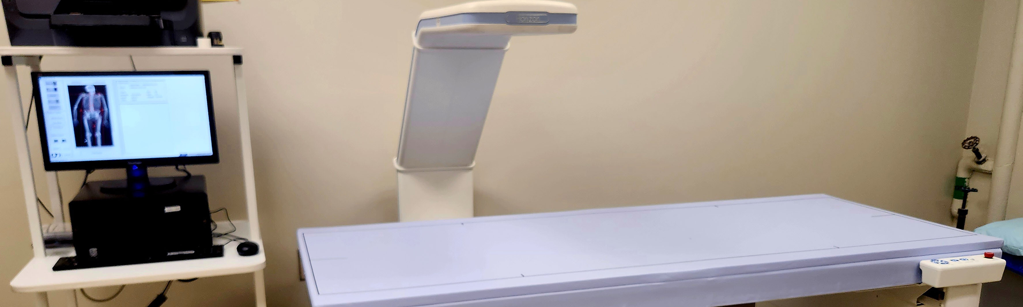
- Dual-energy X-ray absorptiometry (DXA) uses small amounts of X-ray radiation at two different energies to provide very accurate assessments of bone health and body composition. Bone health assessments using DXA are considered the 'gold standard' for bone density determinations.
- Peripheral quantitative computed tomography (pQCT) uses small amounts of X-ray radiation to take cross-sectional images of the arms and legs. These images are used to assess bone health and muscle size in the extremities. pQCT has advantages over DXA as it: (1) assesses bone in three dimensions allowing for true measures of density; (2) provides accurate measures of bone and muscle size, and; (3) separate bones into different compartments (cortical vs. trabecular).
Comprehensive Lung Function:
- Spirometry is used to measure forced expiratory flow rates and volumes.
- Diffusion Capacity of carbon monoxide (DLCO) is a measurement to assess the lungs' ability to transfer gas from inspired air to the bloodstream.
- A Fraction of Exhaled Nitric Oxide (FeNO) test measures the amount of nitric oxide that is exhaled from a breath. Increased levels of nitric oxide are associated with swelling of lung airways and inflammation.
- Impulse Oscillometry (IOS) measures the mechanical properties of the respiratory system (upper and intrathoracic airways, lung tissue and chest wall) during quiet tidal breathing, by the application of an oscillating pressure signal at the mouth.
- Lung Clearance Index (LCI) is measured by multiple breath washout (MBW) is a measure of ventilation in homogeneity and has been shown to be a sensitive lung function test in early lung disease. It reflects disease progression, correlating well with Forced Expiratory volume (FEV1), although it is abnormal at an earlier stage in the disease.
Cardiovascular Health:
- The Flow-mediated dilation (FMD) test is a non-invasive assessment of vascular endothelial function or artery health. Poor artery health is an independent risk factor for cardiovascular disease risk, the number one cause of death in the United States.
- The Microvascular function (MVF) test assesses cutaneous vascular function, an index of microvascular function. Using Laser Doppler Speckle Imaging (LDSI) and laser doppler flowmetry (LDF) cutaneous vascular flux is determined during reactive hyperemia, heat, and various local drug deliveries during iontophoresis and microdialysis.
- The Endothelial independent vasodilation test is a way to assess the vascular smooth muscle function and the ability of the artery to dilate independent of the endothelium. Brachial artery blood flow and arterial diameter are determined at baseline and up to 10 minutes following administration of nitroglycerine.
- Heart Rate Variability (HRV) is a test that can assess the contribution of the parasympathetic and sympathetic nervous system. An EKG is used to determine the variation in time between each heartbeat.
- Carotid Artery Intima-media thickness (cIMT) test measures the thickness of the carotid artery walls, the main artery that feeds the brain. The thickness of the artery in the neck has been used for decades as a clinical tool to measure cardiovascular disease progression and this test can identify local plaque accumulations.
- Arterial stiffness (PWA/PWV) is assessed in humans through the measurement of pulse wave velocity (PWV). The PWV measurement is considered the “gold standard” assessment for measuring arterial stiffness non-invasively. PWV is measured by recording high-fidelity pressure waveforms by placing a tonometer on the carotid, radial, and femoral arteries. The time delay between one arterial site and the arrival of the pressure wave at a 2nd site is calculated, and body surface distance between the respective sites are used to calculate PWV (PWV=distance/time). Using the carotid and femoral sites for PWV is considered a valid measurement of aortic stiffness, which has been demonstrated in numerous studies to be a robust independent predictor of cardiovascular disease risk in adults. Carotid-radial (arm) and femoral-dorsalis (leg) PWV are assessments of peripheral artery stiffness and can also be performed, although the clinical significance of these measurements remains unclear.
Metabolic/Exercise Testing
- Cardiopulmonary Exercise Testing (CPET) is the “gold standard” assessment of exercise capacity is through VO2max testing. There is convincing evidence to indicate an inverse relationship between VO2max and cardiovascular risk, which means that the higher your exercise capacity, the less risk for cardiovascular diseases. During the maximal exercise capacity test, all expired air will be collected through a mouthpiece and analyzed to determine the VO2, or exercise capacity. Heart rate, blood pressure, 12-lead EKG, and rating of perceived exertion (Borg RPE scale 6-20) will be monitored throughout the test in all subjects.
- Resting Metabolic Rate (RMR) is the total number of calories burned when your body is completely at rest. RMR supports breathing, circulating blood, organ functions, and basic neurological functions.
- Six Minute Walk Test (6MWT) is a sub-maximal exercise test used to assess aerobic capacity and endurance. The distance covered over a time of 6 minutes is used as the outcome by which to compare changes in performance capacity. It evaluates the functional capacity of the individual and it provides valuable information regarding all the systems during physical activity, including pulmonary and cardiovascular systems, blood circulation, neuromuscular units, body metabolism, and peripheral circulation.
- Fibroscan® – Transient Elastography is a non-invasive method for measuring liver stiffness. Vibrations of mild amplitude and low frequency are transmitted from the vibrator to the tissues via the transducer, thereby inducing an elastic shear wave that propagates through the tissue. In the meantime, pulse-echo ultrasound acquisitions allow the propagation of the shear wave to be followed and its velocity to be measured, as these are directly related to tissue stiffness: the stiffer the tissue, the faster the shear wave is propagated.