Biorepository
The Georgia Cancer Center Biorepository (GCCB) provides a wide range of professional collection, annotation, and storage solutions. This Biorepository is also home to a state-wide resource, the Biorepository Alliance of Georgia for Oncology (BRAG-Onc), which functions to collect samples from 6 participating sites across the State to represent the diversity of cancer patients within the State and to enhance cancer research in Georgia.
Over the last 4 years, the Biorepository and BRAG-Onc have provided specimens for over 30 individual research projects and have also supplied samples to The Cancer Genome Atlas (TCGA) project. The NHGRI and NCI launched the full-scale landmark TCGA program, with the goal to perform in-depth molecular analysis and sequencing of 4,000 cancer patients representing the 20 most common cancers. Inclusion in the TCGA program earmarks BRAG-Onc and the GCC Biorepository as meeting the most stringent quality control standards for specimen processing.
The Biorepository collects and stores specimens under standardized conditions with accompanying clinical and demographic information and appropriate patient consent, in compliance with the most current Best Practices for Biorepositories as recommended by NCI’s OBBR and ISBER.
Tissue specimens are fast-frozen in vapor phase liquid nitrogen to preserve their integrity and stored in large cryofreezers at -170 to -190°C. Blood is routinely separated into serum or plasma and buffy coats prior to freezing. Blood derivatives are stored at -70 to -80°C. The freezers are monitored constantly and are connected to an alarm system to ensure that the low temperatures are maintained.
Other methods of preparation and handling (such as fresh tissue or tumor cells for culture) are available by arrangement. A specialized Bone Marrow Repository for hematopoietic malignancies and disorders preserves frozen viable mononuclear cells, enriched by gradient centrifugation. There is also access to normal blood products through our collaboration with the Shepeard Community Blood Center.
The repository acts as a critical bridge between the clinical and research endeavors in advancing personalized medicine.
Contact Us
Biorepository
Health Sciences Campus
GCC - M. Bert Storey Research Building
1410 Laney Walker Blvd., CORE 1191, 1190
Rajeev Singh, MBBS, MD, MBA -
Director
706-721-3037
Monday - Friday
8:30 a.m. - 4:30 p.m.
Equipment
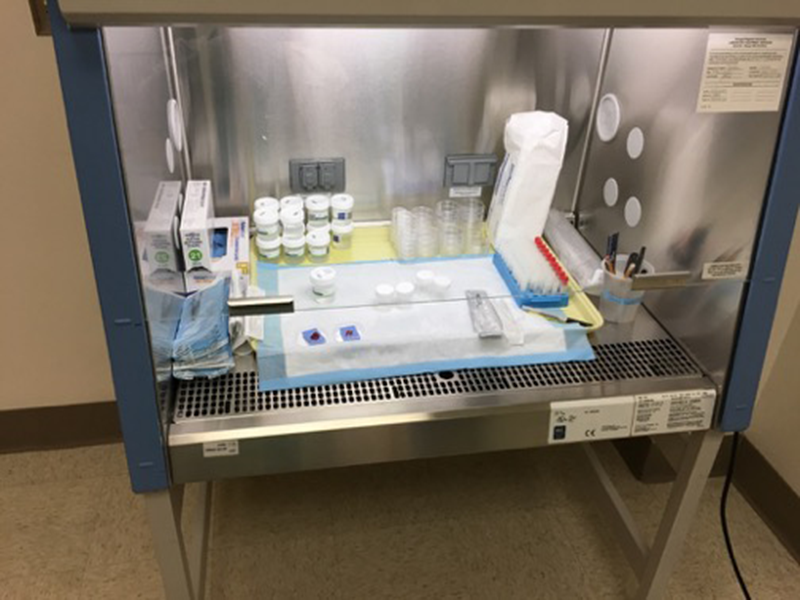
Biosafety cabinet
To maintain safety and sterility, samples are handled in a laminar flow biosafety cabinet in the main laboratory, frozen section room and in the ancillary COVID-19 laboratory.
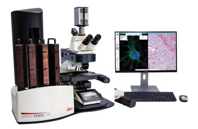
Leica Aperio Versa
Digital slide scanning is carried out using the Leica Aperio Versa. The wide range of objective lenses and motorized nose piece for automatic changing, enables scanning of slides at their ideal resolution. Slides can be scanned anywhere from 0.9mm to 1.2mm thickness, with scan times of 206 secs for 15x15mm at 20x. With the 200-slide robotic slide loader, up to 200 slides may be scanned in one session. Scanned images of appropriate resolution are uploaded to Augusta University Box for secure access and storage. Files in .scn format may be viewed using Aperio ImageScope software from Leica.
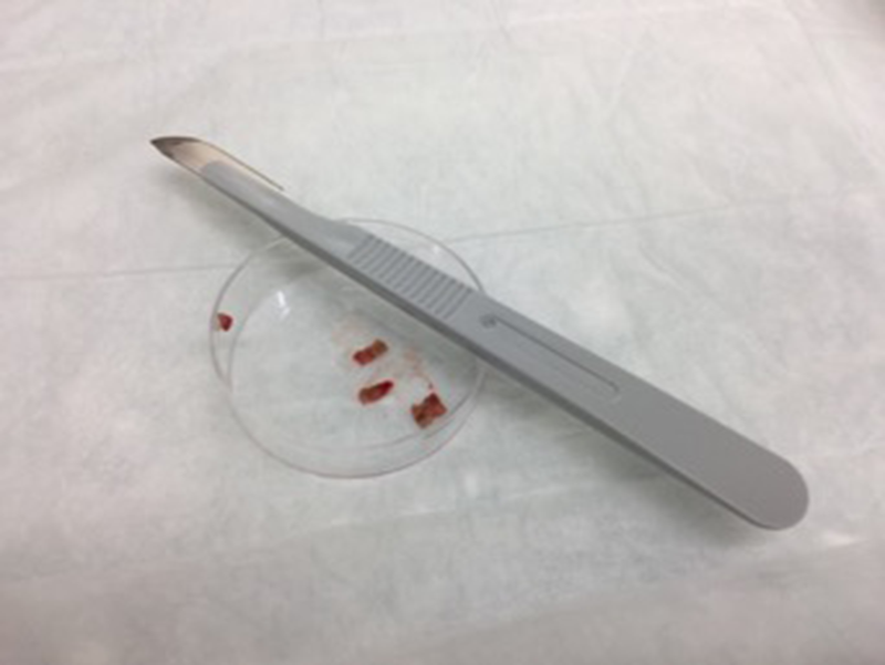
Tissue Aliquots
Surgical samples transported directly from the OR are reviewed by a pathologists’ assistant or by a resident pathologist and ensuring specimen integrity for diagnosis, suitable samples are dissected and provided to biorepository personnel. These samples are annotated and further dissected to provide multiple aliquots that can be flash-frozen or fixed in formalin for subsequent processing and distribution for translational research.
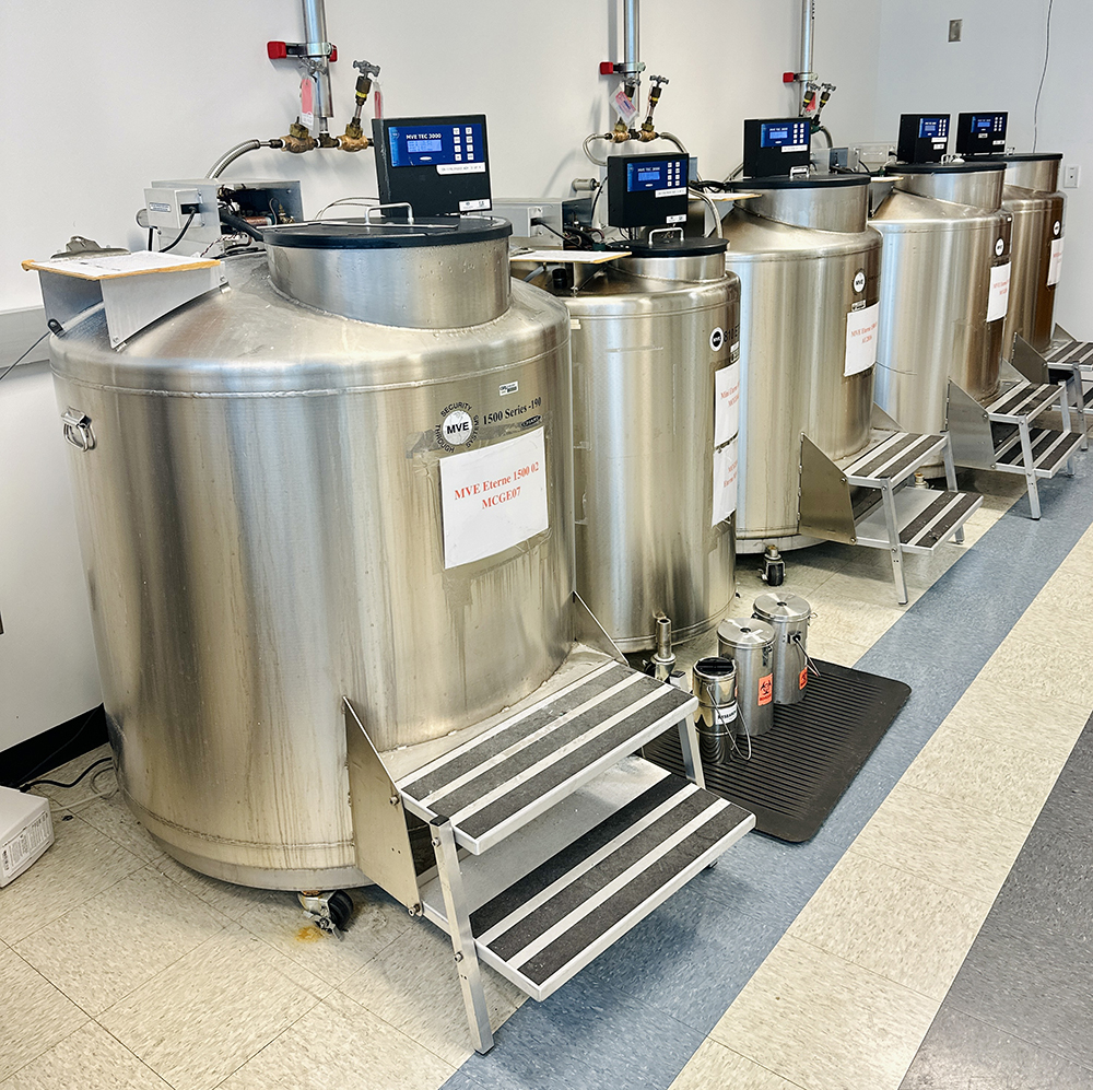
MVE Liquid Nitrogen Freezer Bank
The MVE Series of cryogenic freezers is perfect for the Life Sciences. With enough storage capacity for up to 39,000 1.2/2.0 ml vials, there’s plenty of room for your valuable samples. And with MVE’s 60 years of expertise in manufacturing open-top models like this, you’ll have complete peace of mind that your precious contents will remain viable and protected.
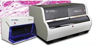
TMA GrandMaster
The TMA Grandmaster is a fully automated tissue microarray production platform with high capacity and high throughput. TMAs are produced by transfer of small tissue cores from a single patient block into a ‘recipient’ block with multiple arrays in parallel. Once generated, multiple tissues are arrayed on a single paraffin block and can be reproducibly interrogated with immunohistochemistry or in situ technologies. The construction of conventional TMAs is labor intensive, imprecise, and time-consuming. The use of an automated tissue microarray system such as the TMA Grand Master is a state-of-the-art solution to improve efficiency, reproducibility and quality. The TMA Grandmaster allows size and spatial flexibility with core diameters ranging from 0.6 mm to 2.0 mm, automated block image control and automated PCR extraction functions.
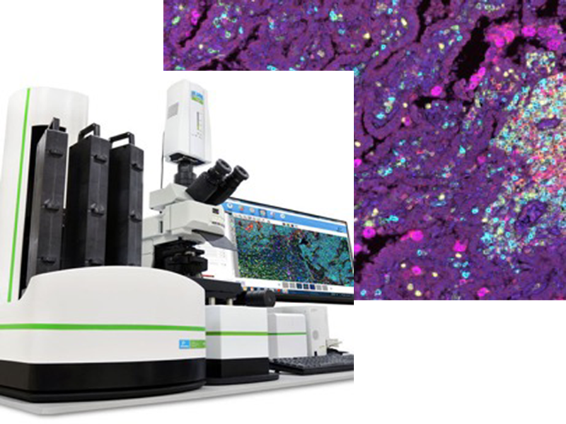
Vectra 3
The Vectra 3 Multispectral Imaging system is an automated brightfield and fluorescence digital scanning microscope. Vectra’s automated, high-throughput imaging capabilities make it an ideal system to extract proteomic and morphometric information from intact tissue sections. The Vectra provides automated slide-handling via a 200-slide robotic slide-loader, brightfield and multispectral imaging technology and pattern recognition-based image analysis software. The system can identify nuclei with DAPI staining and superimpose up to 6 additional fluorophore-conjugated markers. Software algorithms can detect intensity and spatial information to quantitatively evaluate the tissue microenvironment. The Vectra3 is ideal for pre-clinical and clinical trial assessment.
Meet Our Team
Rajeev Singh, MBBS, MD, MBA
- Director, Georgia Cancer Center Tumor Bank
706-721-3037