Selner's Rhinolaryngoscopy Online
John C. Selner, MD
William K. Dolen, MD
Bryan Spofford, MD
Jerald W. Koepke, MD
Chapter 1. UPPER AIRWAY DIAGNOSIS
Routine examination of the upper airway usually consists of inspection of the anterior nares with a nasal speculum and examination of the pharynx with a tongue depressor. In examining the nose, it is essential to use a bright, focused light source and adequate vasoconstriction. Many physicians use an otoscope as a light source; this instrument has a focal length of approximately 2.5 cm with a depth of focus of only 1 cm, permitting examination of only the proximal portion of the nasal cavity. The standard otolaryngologist's head mirror, with a focal length of 10-12 inches, allows the examiner to focus a pinpoint beam of light on the structures to be examined. By skillful manipulation of the patient's head, it is possible to examine large portions of the nasal cavity as well as part of the nasopharynx. Use of a tongue depressor permits evaluation of parts of the posterior pharyngeal wall, but an indirect mirror examination allows a more complete inspection of the nasopharynx, hypopharynx, and the glottic structures. An experienced examiner can evaluate many children and most adults without use of topical anesthesia or sedation. In practice, however, this method has remained unsatisfactory for many situations, and the physician is afforded at best a fleeting glimpse of these important structures. Even in the best of conditions, these conventional methods do not permit examination of the recessed structures of the upper airway, such as the sinus ostia, sphenoethmoidal recess, and eustachian tube ostium.
UPPER AIRWAY ENDOSCOPY
The optically excellent Hopkins nasal endoscopes permit extensive examination of the nose and portions of the nasopharynx, as well as high quality photography of the structures examined. Laryngeal examination is more difficult The cost of a set of these rigid endoscopes with various viewing angles and the relative difficulty in learning to use them has limited their use.
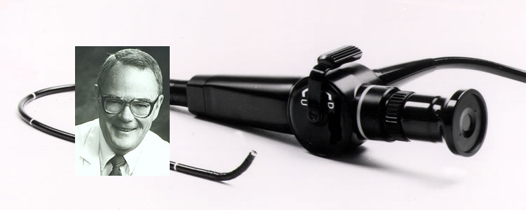
Fiberoptic rhinoscopy is a convenient, relatively inexpensive, and safe alternative. The fiberoptic rhinoscope permits quick and thorough examination of most areas of the upper airway without discomfort to the patient Abnormalities of the septum, turbinates, mucosa, nasopharynx, adenoids, eustachian tube orifice, tonsils, posterior tongue, epiglottis, glottis, and vocal cords can be easily seen. The origin and course of nasal polyps can be identified, and the effect of treatment can be followed. Examinations may be videotaped for permanent record keeping, and videotapes may be replayed and explained to patients.
OTHER DIAGNOSTIC METHODS
Ultrasonic examination of the sinus cavities has enjoyed limited success. Plane x-rays are useful for examination of the sinuses and bony structures of the head, but give limited useful information about the soft tissues of the upper airway, and are occasionally inaccurate in the diagnosis of sinusitis. Computed tomography (CT) provides more detailed images of the bones and soft tissues, and is currently the definitive method for evaluation of the sinuses for inflammatory mucosal thickening. It is expensive, time consuming, and inconvenient. Magnetic resonance imaging (MRI) also provides high resolution images, and has the additional advantage of not employing ionizing radiation. None of these methods is practically useful for routine examination.
INDICATIONS FOR EXAMINATION OF THE UPPER AIRWAY
Indications for Examination
General
- any symptom or historical complaint referable to the upper airway
Nose and Nasopharynx
- nasal obstruction (particularly if unilateral)
- headaches
- facial pain
- epistaxis
- rhinorrhea
- sinusitis
- earache, recurrent or chronic otitis media
- regional adenopathy
- assess result of surgical intervention
Hypopharynx and Larynx
- dysphagia or globus
- hoarseness, other changes in voice quality
- chronic cough
- atypical asthma (laryngeal dysfunction)
Virtually any symptom or historical complaint referable to the upper airway could be an indication for fiberoptic examination. The information obtained will almost always directly influence interpretation of patient complaints, establishment of a diagnosis, and selection of treatment strategies.
Nose
Complaints which might prompt fiberoptic examination of the nose include nasal obstruction (particularly if unilateral), headaches, facial pain, epistaxis, rhinorrhea, sinusitis, earache, recurrent or chronic otitis media, and regional adenopathy. One may also assess the result of surgical intervention.
Nasopharynx
Many common complaints originate in the nasopharynx. These can result in posterior nasal obstruction, anterior facial pain, headaches, epistaxis, rhinorrhea, ear pain, recurrent sinusitis, recurrent otitis media, and regional adenopathy, particularly of the posterior cervical lymph nodes. Examination of the nasopharynx is essential in evaluating these complaints, particularly in patients with nasal obstruction and eustachian-tube dysfunction.
Hypopharynx and larynx
Patients with dysphagia and globus may have distinct pathology of the hypopharynx and larynx. Hoarseness or changes in voice quality may signal pathology in these areas. Gastroesophageal reflux may produce chronic inflammation of the airway resulting in chronic cough and painful swallowing. Patients with upper airway obstruction may present with a history of atypical asthma refractory to usual treatment.
RESEARCH APPLICATIONS
The rhinoscope permits direct visualization of the nasal mucosa. When examinations are videotaped or photographed, results of therapeutic intervention may be compared before and after therapy. A quantitative modification of the basic rhinoscopic technique permits measurement of cross sectional nasal airway in the anterior nose, resulting in the ability-to estimate nasal patency. The patient is seated in an examining chair. The rhinoscope is placed into the anterior nares at a point at which the floor of the nose, the anterior portion of the inferior turbinate, and the nasal airway are in the same field The distance from the nasal meatus to the tip of the rhinoscope is measured, and the image of the nasal airway is videotaped. The videotape is subsequently played back, and its perimeter, defined by the visualized boundaries of the patent portion of the airway, is traced on a transparent plastic sheet. The cross sectional nasal airway area is later measured from the plastic sheet with a computer planimeter. We have used this method to assess the effect of antihistamine and decongestant therapy and for performing nasal methacholine challenges. Its precision is similar to that of anterior rhinomanometry.
In memoriam, John Canty Selner, MD (1936-2006)