This section is an intermediate explanation of how your ears and your brain work together to hear and make sense of sound. By the end of this section you should be able to:
For an introduction to senses and hearing visit: Grades K-5.
Your body has the ability to detect the environment around you from your various senses; sight, hearing, taste, smell, and touch each give you different bits of information that your brain can use to understand where you are and what is going on around you. Hearing is detecting sound, but it is also much more; hearing involves the ability to make sense of those sounds.
In order to hear sounds and understand them several things must happen: First, sound reaches your ear, then that sound is changed into an electrochemical signal and that signal has to travel to your brain. Once your brain receives a signal from your ear, it must be directed to the correct part of the brain that can identify that specific sound. In order for your brain to identify a sound and understand what it means, it must first learn about and be able to recognize that specific sound.
To understand how sound is carried through the ear, you need to understand some of the anatomy. The figure below shows the different parts of the ear:
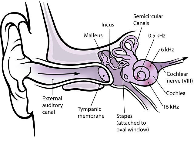
Let’s examine what the different parts of the ear are and what they do. Your ear is more than just what is on the side of your head; that’s called the auricle and it helps guide sound waves into your external auditory canal, or ear canal. At the end of your ear canal is your eardrum, or tympanic membrane; it converts the vibration of air (sound waves) into movement of very small bones in your ear called ossicles. These ossicles consist of 3 bones: the malleus, incus, and stapes. As these bones move, they transmit movement of the eardrum to the oval window, an opening in the cochlea. The cochlea is a spiral structure shaped like a snail’s shell; the hollow inside of the cochlea forms a long winding tube filled with specialized cells called “hair cells”. These hair cells move with sound vibrations and change that movement into electrical impulses. These impulses are carried along the cochlear nerve into the brain and eventually arrive at the auditory cortex. We’ll learn more about that in a moment, but first let’s talk about sound frequencies and frequency discrimination.
Humans can hear sounds in the range of 20 to 20,000 Hz (Hertz). To put it simply, the frequency of a soundwave is how high or low the tone of a sound is. An example of a low frequency sound is the sound of a bass drum, which can produce frequencies in the range of 15 to 100 Hz. On the other hand, when birds sing they produce high frequency sounds in the range of 1,000 to 8,000 Hz. If you are familiar with musical notes, the frequency of C4 (Middle C) is 261.6 Hz and the frequency of A4 (which is higher) is 440 Hz.
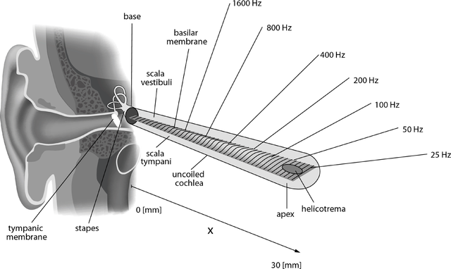
Because the cochlea forms a spiral that gets narrower as it turns, it is able to separate sound into different frequencies. Different parts of the cochlea can detect different frequencies; signals from these different parts of the cochlea go to different parts of the auditory cortex in the brain. If you could unroll the cochlea and stretch it out into a straight line, you would see how the frequency detection range goes from high frequency to low frequency, as shown in the figure below.
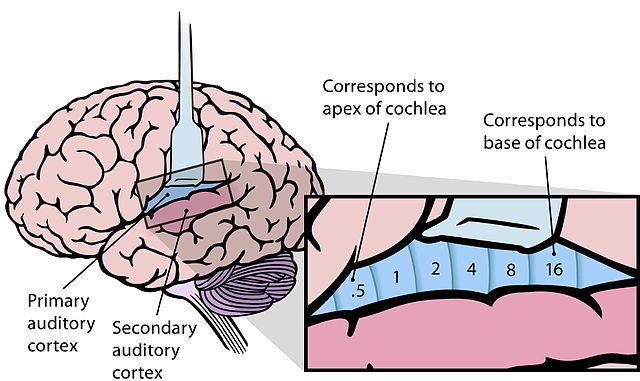
When the signals from these different frequencies reach the brain, they are sent to different areas of the auditory cortex organized by frequency. This forms a sort of “map” of which parts of the brain will be used to hear different sounds, as shown in the picture below. This ability to tell one frequency from another is called frequency discrimination.
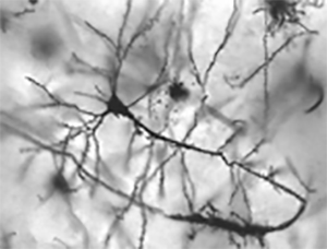
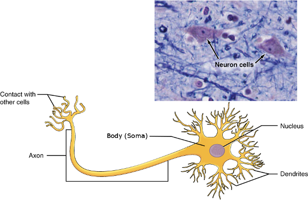
Neurons are cells in the body that transmit electrochemical signals between different parts of the body. Neurons in the brain are often called “brain cells”, while neurons outside the brain are usually referred to as “nerves”. While different neurons serve different functions, their structure and how they work are the same. The figure below shows the anatomy of a neuron.
The body, or soma, of a neuron is the largest part of the cell containing the nucleus and the organelles. Branching out from the soma are dendrites, which receive connections from other neurons. Finally, the axon is the long, thin, part of the neuron that connects to other cells, allowing the neuron to transmit signals to other neurons as well as muscles and some other types of specialized cells.
The point where two neurons connect to each other is called a synapse. Synapses allow two neurons communicate with each other by sending electrical and chemical signals from the axon of one neuron to a dendrite of another neuron. An example of a type of synapse is shown in the figure below.
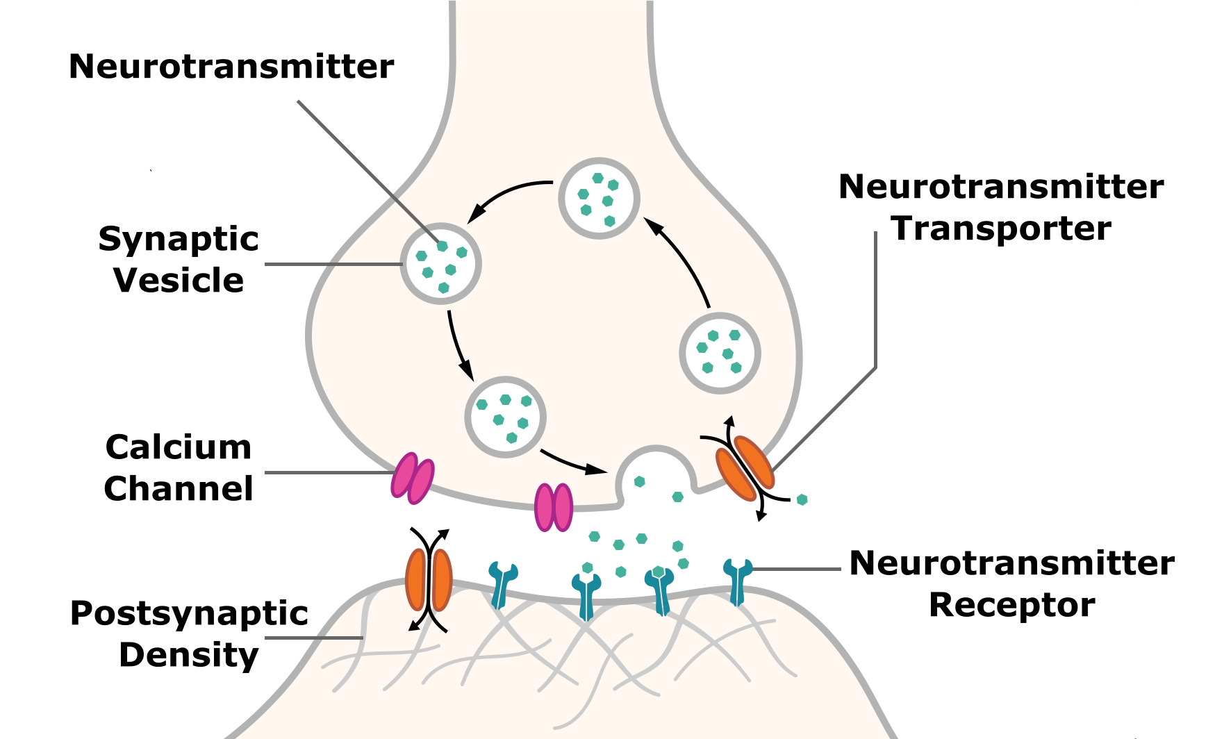
Synapses allow different neurons in the brain to work together. Synapses are capable of changing how they connect two neurons, this change is called plasticity. Synaptic plasticity allows for physical changes in your brain when you learn and form new memories. As you learn, the way your brain cells are connected changes; some new connections are made and some existing connections change to become stronger or weaker. This allows your brain to identify familiar things, such as sounds, with increasing accuracy and precision as you learn. To tell the difference between two different frequencies, groups of neurons in your brain have to learn to respond to only one frequency or range of frequencies, but not to other frequencies.
Here’s an analogy: imagine that the auditory cortex in your brain is a piano player sitting at a piano. When a specific sound, a musical note, reaches the ear and a signal is sent to the brain, the brain must learn to play that note. The brain has to learn which key to play, and also which keys not to play on either side of it. Groups of cells in the brain that are tuned to one target frequency learn to respond to that frequency while groups of cells tuned to other frequencies just above and below that frequency must learn to suppress their response to the target frequency. Synaptic plasticity allows these brain cells to learn how to discriminate between different frequencies by strengthening and weakening connections between these neurons.
Let’s review how your brain hears. First, sound travels to your ear and is transmitted to the cochlea, which is able to separate the sound into different frequencies before converting sound into an electrical impulse. Neurons carry the impulse from the cochlea to the auditory cortex of your brain. Different frequencies detected by the ear are mapped onto specific areas of auditory cortex tuned to those particular frequencies. The brain is learns to discriminate between different frequencies because of a process called synaptic plasticity that allows connections between neurons to change with experience.