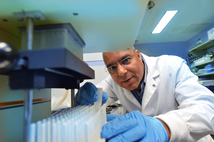
The Georgia Cancer Center supports shared research resources and facilities that provide important support to members of the Cancer Center and their collaborators.
The various resources offer access to state-of-the-art technology and computational support at an affordable cost. Training, consultation and support are also available. The Directors of the various cores are experts in the technologies that are offered in their respective Shared Resources.
Acknowledgment of all GCC Shared Resources is mandatory on publications resulting from consultation with personnel of the Shared Resouces. Significant involvement in the project, significant involvement in the interpretation of scientific data, involvement in the actual writing of a paper, use of original techniques, and/or novel experimental design by the facility staff are required to be acknowledged by co-authorship and acknowledgment of shared resource facility in accordance with all criteria set forth in Augusta University's hare policy on Authorship of Scholarly Activities. Decisions regarding authorship or acknowledgment should take place during the initial phase of the consultation process.

BIOSTATISTICS & BIOINFORMATICS
The Georgia Cancer Center's Bioinformatics Shared Resource offers state of the art bioinformatic support for analyses of genomic data and statistical support. In addition, the Resource manages the Georgia Cancer Center HPC for advanced computing.
BIOSTATISTICS & BIOINFORMATICS ADDITIONAL INFO
Grant Support Provided
The Biostatistics Core (BC) provides in-depth biostatistical review of Cancer Research Center grants, providing important feedback regarding feasibility and statistical considerations in the proposals to both the review panels and investigators. Through collaboration with the BC core in the development of grants and protocols, investigators quickly learn to value the expertise provided by our biostatisticians.
The strongest grants are those where a biostatistician has been involved from the beginning in the design. In general, the likelihood of funding is improved when a biostatistician has fully participated in the development of the proposal. We do not allow a biostatistician’s name to be placed on a grant unless they have been involved in its development or reviewed the grant application.
To ensure the achievements of specific aims, biostatisticians provide critical design and methods support by:
Timing of Grant Support
Biostatisticians that are involved from the beginning need sufficient time to develop a strong grant for submission. The amount of time needed depends on the size of the grant and the agreed upon effort of the biostatistician. The earlier the support request, the more thorough and effective the level of biostatistical support will be, which in turn will increase the ability for your grant to impress critical reviewers.
Investigators should meet with the BC to discuss aims, measurements, criteria for success, and power and prior to any statistics work. Without full understanding of the work, no one can guarantee the quality of outcomes.
Percent Effort
Our policy is that the FTE (full time equivalent) should match the actual effort. Our minimum percent effort on a grant is 5%, although this percentage is rarely enough to cover the scope of the work in a typical grant. For applicable grants, time spent by the biostatistician preparing the grant itself is covered under the cancer center mechanisms.
Manuscript Preparation
Sufficient time is needed for the biostatistician to write or review the statistical methods and the results section of a manuscript or abstract. Whether the biostatistician writes or reviews these sections in a manuscript is determined by the level of collaboration. Thus, communication between the project investigator and the biostatistician is essential in order to meet specific deadlines. Generally two weeks are required for the biostatistician to write their section of the manuscript; however, four weeks may be needed for more complex analyses. Regardless, final versions of manuscripts and abstracts must be reviewed and approved before submission.
Occasionally there is concern about whether or not the biostatistician should be a co-author on the manuscript. It is our policy that the biostatistician (both Ph.D. and M.S.) should be listed as a co-author if there has been a scientific or an intellectual contribution to the research and not be listed in the acknowledgments. If this is going to be an issue with the project investigator, then an agreement with the biostatistician on authorship of any resulting manuscripts should be decided during the initial phases of the project.
Initiating Support
Protocol design is an intensive collaborative work in which biostatisticians play a major role.
BC has the best in-house expertise of biostatistics support for trial design, protocol development, regulatory issues, analysis and reporting. Good protocol or proposal development takes in average of two months. The BC is not responsible for faulty design, methodology if the investigator is not willing to meet with the BC to discuss.
Residents
For Investigator initiated Clinical Trials
For Grant application by Investigators
For other consultation
Booking of server time is done through ilab. iLab allows researchers, PIs, financial managers, and core managers to ensure that they are using valid payment information (Chart Field Combination or CFC) at each step of the request and billing process for core facilities. Financial managers assign CFCs to lab members who will order services from AU cores. Researchers can order services with valid CFC, and core managers can bill for these services knowing that they are using valid CFCs.

BIOREPOSITORY
The Georgia Cancer Center's Biorepository provides a wide range of professional sample collection, annotation, and storage solutions. This Biorepository is also home to a state-wide resource, the Biorepository Alliance of Georgia for Oncology (BRAG-Onc), which functions to collect samples from 6 participating sites across the State to represent the diversity of cancer patients within the State and to enhance cancer research in Georgia.
BIOREPOSITORY ADDITIONAL INFO
A Collaborative Effort
The collection of specimens, coordinated by the tumor bank, requires the collaboration of many individuals, such as surgical oncologists, surgery staff and pathologists. Most important in this process are the patients who donate specimens for future research. It is an opportunity for patients to contribute to science that may lead to better and earlier cancer detection and treatments. And donating makes use of tissue or other material that is unneeded for diagnosis and that would otherwise be discarded.
Available Specimens
The repository collection includes a variety of specimen types, such as tumor tissue and cells, blood and other biofluids as well as normal specimens used as controls. The quality of all specimens is reviewed by a pathologist, and the information is captured in the tumor bank’s database. An imaging system will be used in the future to capture this information.
Human-derived specimens are a very precious resource, and therefore the operations of the tumor bank are overseen by an advisory board, representing Augusta University and other stakeholders, to ensure that the specimens are properly utilized and the privacy of specimen donors is protected.
Biorepository Alliance of Georgia (BRAG-Onc)
The mission of the Biorepository Alliance of Georgia (BRAG-Onc) is to provide a centralized service for biospecimen procurement and distribution across the State of Georgia to advance cancer research.
BRAG-Onc was established to represent the diversity of the cancer patient population in Georgia and to enhance cancer research in the state. The repository collects and stores specimens under standardized conditions with accompanying clinical and demographic information. The collection is supported by a web-accessible database for inventory management and annotation, and a long-term storage facility with backups of cryopreserved specimens. The repository serves as the central coordinating center for the statewide network.
A few representative studies are described below to demonstrate the effectiveness of the Alliance in supporting basic and translational research. Over the last four years, the Biorepository has supported over 30 individual projects.

Acute Myelogenous Leukemia
John Cowell, PhD, FRCPath is the Director of the Molecular Oncology and Biomarker Program and Associate Director for Basic Research at the Georgia Cancer Center. His lab has been a leader in understanding the progression of leukemic stem cells to acute myelogenous leukemia (AML). Samples obtained from the biorepository have helped understand the molecular etiology of AML in specific genetic subtypes and provides opportunities to develop novel treatment regimens.
Targeted glycoproteomic identification of biomarkers for human breast carcinoma
Dr. Michael Pierce, Director of the University of Georgia Cancer Center, has also been using tissues from the Biorepository to study the role of glycans in cancer. Research by Pierce and others has shown that changes in cell glycans can herald the presence of cancerous or precancerous cells. Among his research accomplishments, Pierce and his team isolated a specific enzyme known as Gnt-V that is elevated in colorectal and breast cancer cells, as well as other types of cancer. The team is now investigating ways to inhibit Gnt-V in hopes of developing a treatment that will slow the growth of tumors and prevent metastasis.
The Role of Indolamine-2,3 dioxygenase (IDO) in the suppression of Anti-tumor Immune Responses
Cells from the tumor bank have also been used by Dr. David Munn, Professor of Pediatrics, who works on tumor immunology and the molecular mechanisms of immune suppression and tolerance. A major focus of the laboratory is the role of tryptophan metabolism by the enzyme indolamine-2, 3 dioxygenase (IDO). IDO-expressing dendritic cells are studied with an emphasis on how these cells suppress anti-tumor immune responses via IDO and downstream pathways. The laboratory employs clinical and translational strategies designed to enhance antitumor immune responses using IDO-inhibitor drugs.
Heat shock Protein 90 as a Therapeutic Target
The focus in the Chadli laboratory is on the identification of heat shock protein 90 (Hsp90) inhibitors as anti-cancer reagents. The Hsp90 chaperoning machine maintains the conformation and stability of many oncogenic proteins, transcription factors, steroid receptors, metalloproteases, and nitric oxide synthases that are essential for cancer cell survival and proliferation. The Hsp90 machine is therefore an exciting therapeutic target, the inactivation of which would deliver a combinatorial attack on multiple signaling pathways, leading to a more efficient killing of cancer cells and reducing resistance to chemotherapy. His laboratory has developed novel small molecule inhibitors of the Hsp90 chaperoning machine and has used samples from the biorepository to test the efficacy of these new drugs.
Whole Methylome Sequencing in Lymphoid Malignancies
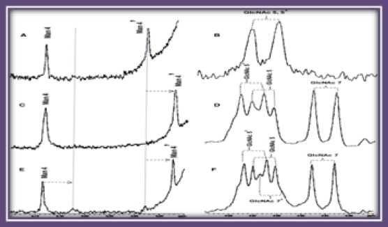
The Shi laboratory has spent a significant amount of effort on developing high-throughput technologies for dissecting complex epigenetic regulation in normal and cancer cells. One of their most recent innovations was the development of a targeted bisulfite sequencing method based on solution-based sequence capture technology. Currently, they are utilizing these high-throughput bisulfite-sequencing methods for profiling DNA methylation patterns in leukemias and lymphomas. They have demonstrated that aberrant epigenetic gene regulation in lymphoid malignancies involves DNA methylation, histone modifications, and spatial conformation of the genome. Samples obtained through the Biorepository have been used to elucidate the epigenetic profiles in chronic lymphocytic leukemia which can now distinguish subtypes that have better outcomes.
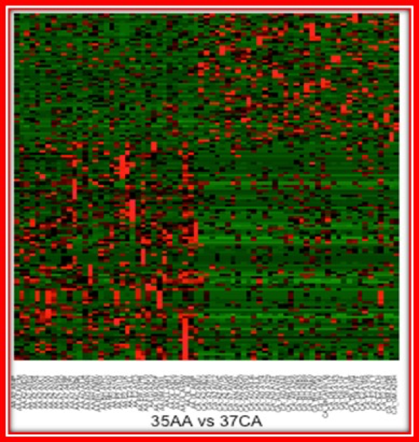
Mutational Analysis of Triple Negative Breast Cancers to Examine Racial Disparities in Outcomes
The Hawthorn Laboratory is in the process of sequencing triple negative breast cancers (TNBCs) obtained from the biorepository. Triple negative breast cancer is defined by the absence of detectable estrogen and progesterone receptor expression levels and by the lack of the epidermal growth factor receptor 2 (HER2) gene amplification. Because it is lacking these tumor-specific molecules there are currently no targeted therapies to treat this type of breast cancer. The survival rate of women developing this breast cancer subtype is a devastating 14% while those who develop estrogen positive tumors have a survival rate of 97%. Women of African descent (AA) have a generally lower rate of breast cancer incidence than women of European descent (CA), however, 20% of tumors in CA patients are TNBCs, while ~40% of breast cancers in AA patients are TNBCs. Dr. Hawthorn is using whole genome sequencing to profile TNBC tumors and examining the profiles of CA and AA patients to determine if the disease is different between the two racial groups. There is currently very limited data on AA TNBC as a separate disease entity so that access to the samples provided by BRAG-Onc are critical to the success of this important study.
The Niacin/Butyrate Receptor GPR109A Suppresses Mammary Tumorigenesis By Inhibiting Cell Survival
Drs. Ganapathy and Thangaraju of the Signaling and Angiogenesis Program within Georgia Cancer Center have studied the tumor suppressor function of a receptor for niacin and butyrate called GPR109A. Using samples received from BRAG-Onc, these investigators found that GPR109A is expressed in normal breast tissue but is silenced in tumor tissue. Studies have shown that activation of this receptor results in the inhibition of genes involved in cell survival of breast cancer cells. These findings have been confirmed in cell lines and mouse models of breast cancer and suggest the GPR109A is a tumor suppressor in the mammary gland. Studies are now underway to determine if therapeutic activation of this gene may be an effective strategy in the treatment of breast cancer.
In addition to the projects presented below, the biorepository has been involved in The Cancer Genome Atlas (TGCA) project. This very important undertaking is currently the National Cancer Institute’s (NCI’s) flagship research program, involving both intramural NCI scientists as well as extramural scientists from leading research centers and universities around the globe. In 2003, the first complete sequencing of the entire human genome was completed at a cost of over $3-billion. Sequencing technology has since exponentially improved, bringing the cost and time required down to remarkable levels in just a decade. In 2009, the NHGRI and NCI launched the full-scale TCGA program, with the goal to perform in-depth molecular analysis and sequencing of 4,000 cancer patients from the 20 most common cancers.
Since 2010, BRAG-Onc has been a vital member of the TCGA network and has contributed specimens from multiple cancer types including Head and Neck cancer, Thyroid cancer, Ovarian cancer, and Urinary Bladder cancer. Inclusion in the TCGA program earmarks BRAG-Onc as meeting the most stringent quality control standards for specimen processing, alongside peer institutions such as MD Anderson, Fox Chase Cancer Institute, Memorial Sloan-Kettering, Imperial College London, and UCSF.
While it will likely take years to fully analyze the wealth of data being produced, the promises of this effort are already being realized. In addition to the NCI grant to fund the specimen collection and annotation, AU Cancer Center physicians Weinberger and Bollag were recently included as co-authors in a major TCGA study published in the journal Nature. Further publications and investigator-initiated grant proposals are also anticipated.
The Georgia Cancer Center Biorepository has amassed a collection exceeding 30,000 biological samples from over 18,000 registered donors. As a centralized shared resource, the facility and expert staff add value through experience, efficiency, standardization, accountability, protection of patient confidentially and timely completion of research.
Our services include:
1.) In order to obtain specimens, users must fill out the Request for Tissue Search to indicate the type and number of samples. The completed form can then be forwarded to Archana Laknaur. The personnel will conduct a search of the Biorepository collection to determine if the required samples are available. In the future, the Biorepository will have a searchable database available for these inquiries.
2.) Once confirmation of available samples has been obtained, requestors are required to fill out the Application for Banked Specimens. Please be as informative as possible when completing the application and include specific details on the scientific merit, statistical power calculations for the number of samples requested, and the planned experimentation. This will ensure timely review of your application. Applications that do not contain sufficient information for reviewers to make an informed decision will be returned.
3.) Institutional Review Board (IRB) approval or an exempt declaration is required for any study involving human tissues. The IRB has sanctioned two main biospecimen collection protocols: one for solid tumors (611107) and one for hematologic malignancies (611132). Beyond these protocols, Investigator-initiated biorepository projects require full IRB submission.
4.) Biorepository personnel will notify you to arrange collection of the samples. Sections and slides are also available for each of the samples upon request. You will be required to sign an acknowledgement upon receipt of tissues.
BRAG-Onc Membership
Here is a look at the current medical centers, clinics, etc. who are members of the Biorepository Alliance of Georgia (BRAG-Onc).
FLOW & MASS CYTOMETRY
The Georgia Cancer Center's Flow & Mass Cytometry Shared Resource provides investigators access to six flow cytometers, including two cell sorters and a variety of analytical instruments.
FLOW & MASS ADDITIONAL INFO
Flow & MAss Cytometry
Useful Links
We have changed our staff-assisted cell sorter rate at $90/hr for both sorters. New pricing is effective from 10/1/2023.
All users of the Georgia Cancer Center Flow Cytometry Shared Resource should acknowledge the facility in all publications resulting from work performed in the resource.
The types of acknowledgment are described below. Cases of disagreement or potential conflict arising from prior agreements (or more likely, lack of prior agreements) will be assessed and resolved by the administration overseeing the Shared Resource and/or office of Senior Vice President for Research.
I have used the identical equipment somewhere else. Do I have to be trained again?
We like to orient everyone to the GCC facility protocols and procedures for the instrumentation
so we like to meet all new users prior to them utilizing the facility on their own.
What type of samples can be run on the flow cytometers?
 Any single cell or single particle suspension can be analyzed, including mammalian
cells, bacteria, yeast, dissociated tissue and polystyrene beads. If your cells or
particles are not in a single cell suspension, you cannot perform flow cytometry.
Cells that are not from whole blood must be filtered. Persons working with adherent
cells must make sure that their cells stay in a single cell suspension before and during the entire process. It is recommended to suspend cells in a product called Accutase,
which can be purchased from its creator, Innovative Cell Technologies, or from many
other sources, e.g., SigmaThermo Fisher, Millipore Sigma, BioLegend. Filtering alone is insufficient to prevent adherent cells from reforming clumps.
Any single cell or single particle suspension can be analyzed, including mammalian
cells, bacteria, yeast, dissociated tissue and polystyrene beads. If your cells or
particles are not in a single cell suspension, you cannot perform flow cytometry.
Cells that are not from whole blood must be filtered. Persons working with adherent
cells must make sure that their cells stay in a single cell suspension before and during the entire process. It is recommended to suspend cells in a product called Accutase,
which can be purchased from its creator, Innovative Cell Technologies, or from many
other sources, e.g., SigmaThermo Fisher, Millipore Sigma, BioLegend. Filtering alone is insufficient to prevent adherent cells from reforming clumps.
What should I bring my samples in?
For analysis, samples should be brought in 12 x 75 mm round bottom style, polystyrene plastic Corning Falcon test tubes (catalog #352008). The same size tubes from other manufacturers may not necessarily fit the sample O-ring on the analyzer flow cytometers. For sorting, samples should be brought in 12 x 75 mm round bottom style, polyproylene or polystyrene plastic Corning Falcon test tubes (catalog #352063 and 352008, respectively) or in 15 ml conical tubes.
I need protocols for sample preparation. Where can I obtain them?
This Web site has a number of links to important and comprehensive protocols. Click on the Protocols & Useful Links section of the facility's Web site to access them.
How many cells do I put into each tube?
For analysis, we recommend that you put 1 million cells in 500μl of FACS buffer into each test tube.
What controls do I need?
There are different types of flow cytometry controls that are necessary every time an experiment is performed. Basic flow cytometry controls consist of negative and single color controls. Negative controls consist of cells alone or an isotype control. Single color controls are used for compensation of fluorochrome emission overlap. Single color controls must have sufficient signal to be able to adjust instrument settings (i.e. they MUST be visibly POSITIVE). Single color controls do not necessarily have to be the same antibody as the antibody of interest as it is the fluorophore that is being controlled for, not the antibody. If the conjugated antibody of interest is not brightly expressed, that antibody would not be an appropriate single color control. Some manufacturers sell beads that are designed to bind antibodies, creating the necessary single color controls that flow cytometry requires (Thermo Fisher OneComp eBeads, UltraComp eBeads and AbC Total Antibody Compensation Beads). Some persons may and should choose to prepare additional controls consisting of fluorescence-minus-one [FMO] controls. FMO controls determine how to analyze the sample, i.e., to assist with gate setting for positive and negative expression. They should be considered a must whenever accurate discrimination is essential or when antigen expression is relatively low. FlowJo's 'Daily Dongle' provides an explanation of FMO vs. Isotype Controls. FMO controls cannot be used to set compensation. Related to typical flow cytometry controls, positive controls are used to ascertain the activity of an antibody of interest and are required in certain situations. For example, you may want to prove that your cells do not express a particular antigen. In this situation, you should supply a different cell type that expresses the antigen to prove that the antibody is specific to the epitope of interest. Experiments run without all of the appropriate controls make the interpretation of the information difficult, and any resulting conclusions must be considered suspect.
Can dye X be run in the facility?
It depends upon the dye's excitation and emission characteristics. The facility is equipped with a variety of lasers although it is not comprehensive. Look over the GCC flow cytometry equipment webpage for a summary table of the available lasers and details of fluorochrome detectors available on each instrument. In addition, check out the fluorescent spectrum viewers from BD Biosciences, Thermo Fisher, BioLegend and Enzo Life Sciences found in the Protocols & Useful Links.
Can dye X and dye Y be run together?
It depends upon the dyes' emission characteristics and the instrument. Again, check out the fluorescent spectrum viewers from BD Biosciences, Thermo Fisher, BioLegend and Enzo Life Sciences found in the Protocols & Useful Links.
I have potentially infectious (biohazard) specimens. What do I do?
The primary shared resource laboratory room (CN4158C) does not currently accommodate any biohazardous specimens (BSL1 level). Specimens must be inactivated of all pathogens before they are brought into the facility. Commonly, 1-2% paraformaldehyde is used. The laboratory recommends liquid methanol-free EM grade 16% paraformaldehyde from Electron Microscopy Sciences. This can be diluted using PBS. (10 x 10ml glass ampules are $22.50). Users should be aware that Invitrogen has a variety of LIVE/DEAD Fixable Kits that use an amine reactive dye that allows users to fix cells and to subsequently discriminate live from dead cells in the original sample. Other than preserving cells, these kits have the added benefit in that they inactivate pathogens. The exception to the BSL1 rule is for the sorter flow cytometer, which is housed in a different area (CN4146C). That instrument and the procedures associated with its operation allow it to be capable of sorting/analyzing specimens at the BLS2+ level. Use of this instrument is currently provided as a service only. Please contact us for details.
I would like to sort cells but I am uncertain how to do that. What do I do?
Persons that have never sorted cells before must first contact facility personnel. They will ascertain the feasibility of your sort goal, provide you with guidelines associated with sorting cells and create an appointment for sorting with you.
How can I reduce nonspecific binding?
This section is taken pretty much intact from the FAQ's for Flow Cytometry Acquisition/Analysis on the University of Virgina's Flow Cytometry Facility's webpage formerly directed by Joanne Lannigan — Thanks, Joanne! Nonspecific binding can be due to several reasons: Too much antibody will increase the amount of nonspecific binding of your negative population, which will reduce the signal-to-noise ratio. You should perform titrations for every antibody you use to determine the optimum concentration. Nonspecific binding can also be due to Fc-receptor binding. Use IgG of the same species as your antibody of interest to block nonspecific binding. Incubate samples with a final concentration of 2 mg/ml of IgG for ~10 minutes at room temperature before adding antibodies. Using monoclonal antibodies specific for Fc receptors to block Fc-mediated binding can also help reduce background binding. The use of directly conjugated antibodies can also reduce the amount of non-specific binding. If your antibody is not commercially available as a directly conjugated antibody, there are a number of simple procedures with which you can easily conjugate your antibody:
How often are data files removed from the computers?
No data files are allowed to be stored on the computers in the lab. After running your samples you should immediately transfer your data files into BOX and subsequently transfer them to your own computer. Your data and your experiments are ultimately your own responsibility.
How to schedule instrument use:
Scheduling for all equipment in the core facility is accomplished using iLab (Agilent CrossLab), a web-based electronic calendar.
How much do the cytometers cost per hour?
A current schedule of the facility's fees can be viewed above. Users will be billed for the time that's actually used.

IMMUNE MONITORING
The Georgia Cancer Center's Immune Monitoring Shared Resource provides comprehensive immune monitoring services and standardized immune-based assays for basic, clinical and translational studies.
IMMUNE MONITORING ADDITIONAL INFO
The IMSR works closely with the Flow Cytometry Resource, the Integrated Genomics Shared Resource, and the Tumor Tissue and Serum Repository at the Georgia Cancer Center.
The Georgia Cancer Center's Immune Monitoring team is proud to offer the services below to all in-house and external researchers looking for certain types of assistance with their data and experiments.
Immune Monitoring Publications
Choudhary, V., Uaratanawong, R., Patel, R.R., Patel, H., Bao, W., Hartney, B., Cohen, E., Chen, X., Zhong, Q., Isales, C.M., et al. (2019). Phosphatidylglycerol Inhibits Toll-Like Receptor–Mediated Inflammation by Danger-Associated Molecular Patterns. J Invest Dermatol 139, 868-877.
Singla, B., Ghoshal, P., Lin, H., Wei, Q., Dong, Z., and Csányi, G. (2018). PKCδ-Mediated Nox2 Activation Promotes Fluid-Phase Pinocytosis of Antigens by Immature Dendritic Cells. Frontiers in Immunology 9, 537-537.
Xiao, W., Klement, J.D., Lu, C., Ibrahim, M.L., and Liu, K. (2018). IFNAR1 Controls Autocrine Type I IFN Regulation of PD-L1 Expression in Myeloid-Derived Suppressor Cells. The Journal of Immunology 201, 264-277.
Sharma, M.D., Rodriguez, P.C., Koehn, B.H., Baban, B., Cui, Y., Guo, G., Shimoda, M., Pacholczyk, R., Shi, H., Lee, E.-J., et al. (2018). Activation of p53 in Immature Myeloid Precursor Cells Controls Differentiation into Ly6c+CD103+ Monocytic Antigen-Presenting Cells in Tumors. Immunity 48, 91-106.e106.
Please see our iLab website for currently available services and detailed information. Click this link to download a spreadsheet listing all of our prices and fees.

INTEGRATED GENOMICS
The Georgia Cancer Center's Integrated Genomics Shared Resource houses a complete Illumina Sequencing Facility, including Illumina HiSeq, NextSeq, MiSeq and an Ion Proton instruments. The Resource also supports Affymetrix array technologies and analytical support.
Please note that pricing is subject to charges of the suppliers.
All services are booked through iLAB
The Integrated Genomics Core is thrilled to launch a Core Lab Grant application in collaboration with 10X Genomics with the intent of supporting AU researchers seeking to generate pilot data using cutting-edge solutions from 10x Genomics for genomic and translational research.
How to apply?
Next Generation Sequencing
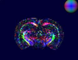
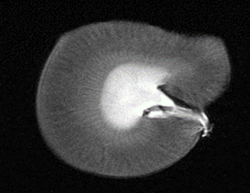
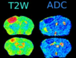
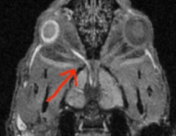
SMALL ANIMAL IMAGING
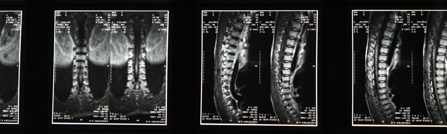
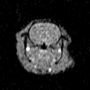
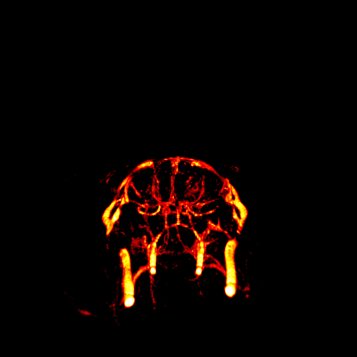
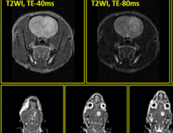
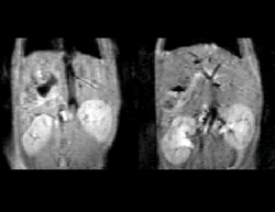
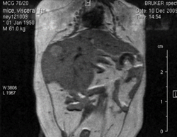
The Georgia Cancer Center's Small Animal Imaging Shared Resource provides magnetic resonance imaging (MRI) for small animals and offers whole-body and other imaging protocols specific to MRI such as quantitative MRI protocols.
SMALL ANIMAL IMAGING ADDITIONAL INFO
The Small Animal Imaging Resource provides a wide range of imaging and radiation treatment resources for animal research. The preclinical services are translational in operation while remaining cutting edge in the advancement of basic science research. Services are available to the Augusta University research enterprise and other outside scientific investigators.
The Small Animal Imaging shared resource takes an orderly approach to managing your imaging project and employs a regimented method for providing Quality Assurance and Control for all image acquisition and image analysis. Small Animal Imaging shared resource incorporates years of experience in research project management and preclinical level image analysis in every imaging project that it is involved for translational studies.
The Small Animal Imaging shared resource provides low-cost imaging services to the Augusta University research community. Prices for imaging, data analysis, and consultation are intended to be competitive with the charges for similar services at other academic institutions.
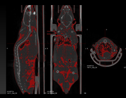
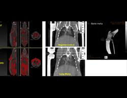
Single photon emission computed tomography (SPECT)
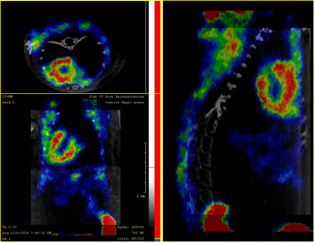
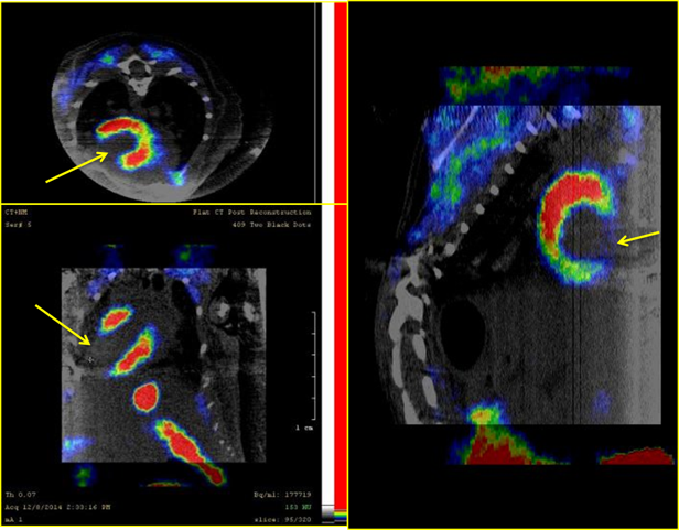
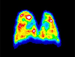
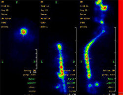
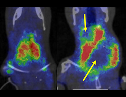
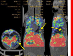
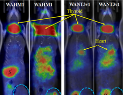
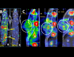
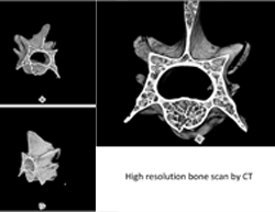
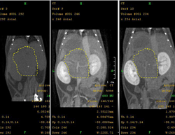
Computed topography
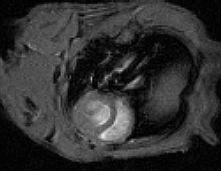
MRI Images
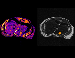
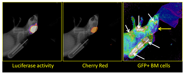
Bioluminescence Imaging/Fluorescence Imaging
Fee & Payment
Small Animal Imaging provides low-cost imaging services to the Augusta University research community.
Prices for imaging, data analysis, and consultation are intended to be competitive with the charges for similar services at other academic institutions. All charged usage will be pro-rated in ½-hour blocks, in accordance to time used. All scheduling and billing are through iLab.
Klaus Ley is exploring the role our immune response plays in atherosclerosis, and his aim is to develop a vaccine and drugs that leverage human immunity to tackle the disease.
For more than two decades, Ley has applied his knowledge of immunology to understand the role that immune cells play in atherosclerosis. White blood cells swim in the blood, helping to protect the body from infection. To do their job, they must adhere to the blood vessel wall. In a chronic disease like atherosclerosis, this happens over and over again, eventually making the lumen narrower and the wall harder. So, instead of helping to solve a problem, the immune cells turn against the body — they actually hasten the atherosclerosis process.
As co-director of the Immunology Center of Georgia, Ley will build on his research while also recruiting candidates to establish a world-class immunology research center focusing on vaccines, cancer immunology and vascular immunology.